
Service
Proteomics Facility
LC-MS Equipment
We are running three quadrupole orbitrap instruments (QExactive, QExactive Plus, Exploris 480) and one linear ion trap (LIT) FT-MS instrument (OrbitrapXL+ETD), that are coupled online via electrospray ionization (ESI) source interfaces to nanoUHPLC (LC-MS). NanoUHPLC reversed phase (RP) separation enables the use of columns with a length of up to 50 cm that in conjunction with sub-2μm bead capillary columns offer superior separation power for complex tryptic peptide mixtures. Recently the EvosepOne HPLC system that coupled to the Exploris 480 can be operated in both the high-throughput microflow and highly sensitive whisper nanoflow (100 nl/min) LC-MS mode was added. We constantly monitor (QC) the LC-MS setup with the help of in-house adopted software (Biognosys QuiC, PTXQC).
Prefractionation and sample preparation

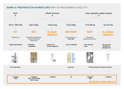
A mammalian cell expresses ~10,000 to 12,000 protein-coding genes with an average protein copy number ranging from 1,000 to 10,000 molecules per cell. Therefore, the number of different proteoforms (due to alternative splicing, alternative transcription and translation start points, PTMs etc.) present in a given cell type is estimated to range between 200,000 to 1,000,000. Currently, even the most advanced LC-MS platform is not able to deal with this complexity and this is why prefractionation is considered key to address this under-sampling problem. Pre-fractionation of high complexity samples (e.g. whole cell proteomes) is commonly achieved either at the protein level by SDS-PAGE or at the peptide level via peptide-SAX, SCX, HpH-RP or RP-SCX (iST fractionation).
Depending on the sample, proteolytic digestions are performed either in-gel, in solution/suspension (iST or SP3 technology), on-bead (affinity pull-downs) or by FASP (filter-aided sample preparation). Sample preparation by SPE (solid phase extraction) is performed offline (semi-automated) in a microcolumn-in-a-tip (STAGE tips) format. This set-up is very flexible and can accommodate reversed phase (standard), strong anion and cation exchange (SAX, SCX), HILIC (hydrophilic interaction chromatography) as well as affinity chromatography beads (e.g. titania-MOC for phosphopeptides).

Data Analysis
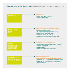
Quantification of SILAC-MS data is achieved with the MaxQuant and Perseus software environment. Similarly, for conducting label-free quantitative proteomics, we make use of the MaxQuant LFQ algorithm followed by R scripts and lima statistics. The bioinformatics pipeline additionally consists of the Thermo Proteome Discoverer 2.5 platform (LFQ, TMT quantification) and a PEAKS Studio workstation connected to a Mascot Server for automated (de novo) peptide sequence tag assisted multi-engine database searching. Likewise, DIA/HRM and targeted PRM data are processed on a Spectronaut Pulsar/SpectroDive workstation.
Core Service
Standard service includes protein ID from silver or colloidal coomassie stained gels, peptide mapping (protein characterization by several proteases), and investigation of simple bead-associated proteomes/interactomes. Deep analyses of high-complexity proteomes (e.g. whole cell and organellar proteomes), studies of PTMs and sophisticated quantitative analyses (large scale interactomics, APEX proximity labeling) are much more time consuming and are therefore considered as scientific collaborations. Single-shot proteome-wide analysis identifying and quantifying proteins in the range of two to five thousand protein groups are available on a routine basis. Users hand-in their samples via our in-house sample submission and archiving system (MS-Web) enabled by a web browser interface:
MS-Web
Research
Our research focus encompasses the application and development of proteomic methodologies for elucidating protein-RNA and protein-DNA interactions. Moreover, spatial proteomics through proximity-labeling approaches (e.g. APEX) as well as the analysis of chromatin and transcription factor proteomes define our key interests. In this context, we pursue targeted PRM analysis of histone PTMs in mouse and human cells.